Tests
Modern spine care has a broad range of tests and diagnostic procedures which may be utilized to understand your condition and establish a treatment plan. Here are a few of the more common tests that may be performed.

X-rays, CT Scans and MRI's provide excellent images of various spinal disorders. However, they cannot show pain. Spinal injections, typically used to control pain, are also used diagnostically to locate the pain source. Diagnostic spinal injections include discography (discogram), selective nerve root block (SNRB), sacroiliac joint injection and facet joint injection.

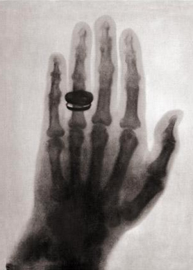
X-rays were discovered in 1895 and have become the most common type of imaging study used by spine specialists to confirm a diagnosis. Through the years the technology has significantly improved. Today, the dose of x-ray needed to produce quality film images is just a fraction of what was required in the past.

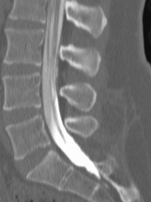
Computerized tomography (CT or CAT scans) combines x-ray science with computer technology to produce more focused and informative images than traditional x-rays. CT scans produce clear pictures of bone, soft tissue, organs, intervertebral discs and the spinal cord. The images can be reproduced in black/gray/white tones or color. To enhance the image, a contrast medium (dye) can be injected into the patient during the test.

When a CT scan and myelography are combined, images are produced that clearly show both the bony structures of the spine and the nerve structures. These images are invaluable to physicians as they diagnose a patient's spine problem.

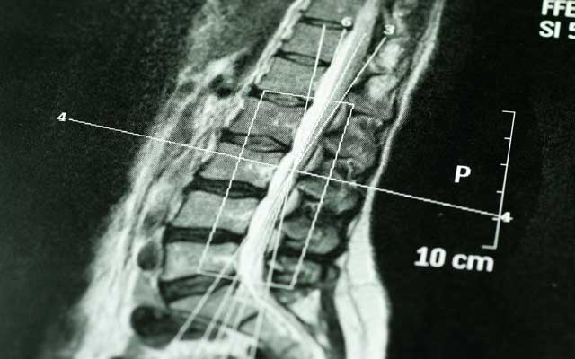
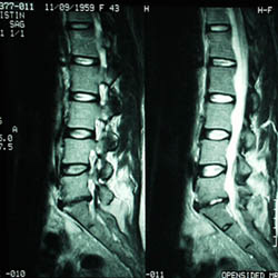
One of the diagnostic tools that we use is Magnetic Resonance Imaging (MRI), which can help us to identify various spinal pathologies including infection, nerve impingement, disc herniation, spinal cord compression, low back and neck pain, and tumor.
